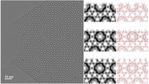

 Metal–organic frameworks (MOFs) are often synthesized using various additives to modulate the crystallization. Here, we report the direct imaging of the crystal surface of MOF MIL-101 synthesized with different additives, using low-dose high-resolution transmission electron microscopy (HRTEM), and identify three distinct surface structures, at subunit cell resolution. We find that the mesoporous cages at the outermost surface of MIL-101 can be opened up by vacuum heating treatment at different temperatures, depending on the MIL-101 samples. We monitor the structural evolution of MIL-101 upon vacuum heating, using in situ X-ray diffraction, and find the results to be in good agreement with HRTEM observations, which leads us to speculate that additives have an influence not only on the surface structure but also on the stability of framework. In addition, we observe solid–solid phase transformation from MIL-101 to MIL-53 taking place in the sample synthesized with hydrofluoric acid.